Home
/ Abdominal Blood Vessels Labeled - Blood Vessel Wikipedia - The aorta is the large artery leaving the heart.
Abdominal Blood Vessels Labeled - Blood Vessel Wikipedia - The aorta is the large artery leaving the heart.
Abdominal Blood Vessels Labeled - Blood Vessel Wikipedia - The aorta is the large artery leaving the heart.. Advertising on our site helps support our mission. Because arteries are moving blood being pumped out by the heart. The identification of abdominal vessels using ultrasound is based on knowledge of their normal location, appearance and relationship to specific organs. Lateral view with the head to the right. That being said, all arterial blood delivered to this region comes via branches of the abdominal aorta, and all venous blood eventually finds its way back to.
Arteries are a type of blood vessel. The venous drainage of the abdomen is carried out by the portal venous system and the systemic venous system. Label the abdominal blood vessels using the hints provided. Efferent branchial arteries injected with red latex. In contrast, veins carry blood back to the heart.
Abdominal Blood Vessels Labeled Arteries Veins Atlas Of Anatomy Hma Practical 3 For Monday July 23 And Wednesday July 25 from i0.wp.com Katy wallis at state college of florida This video series covers the blood vessels for anatomy and physiology ii students. Label the intestinal structures using the hints provided. Efferent branchial arteries injected with red latex. The venous drainage of the abdomen is carried out by the portal venous system and the systemic venous system. They also take waste and carbon dioxide away from the tissues. Nerves, blood vessels, and lymphatics are present throughout. The aorta is the largest blood vessel in the body.
It's the preferred screening method for an abdominal aortic aneurysm, a weakened, bulging spot in the abdominal aorta — the major blood vessel that supplies blood to the body.
Label the biliary passages and associated structures using the hints provided. Label the abdominal blood vessels using the hints provided. We will include an analysis of the normal doppler waveforms of the abdominal vessels. Neurovasculature of the abdominal wall explore study unit superior epigastric artery: Label the biliary passages and associated structures using the hints provided. That being said, all arterial blood delivered to this region comes via branches of the abdominal aorta, and all venous blood eventually finds its way back to. (superficial epigastric visible at upper left.) the left femoral triangle. Blood, the heart and the vessels that carry blood around the body together make up the cardiovascular system. 3 the superficial vessels include the superficial epigastric and the superficial circumflex iliac vessels. The videos are done by dr. The abdominal aorta is the largest blood vessel in the abdomen. An abdominal ultrasound is done to view structures inside the abdomen. It has a number of important relationships and branches, which very commonly appear in exam questions and anatomy spotters.
However, the imaging test may be used to diagnose or rule out many other health conditions. The abdominal aorta enters the abdomen through the diaphragm at the level of the twelfth thoracic vertebre and continues to just below the umbilical area, where it splits into the right and left common iliac arteries. This video series covers the blood vessels for anatomy and physiology ii students. Before lab, read through the procedure and draw diagrams of the blood vessels you will be finding and label these. Abdominal wall anatomy that is clinically pertinent to the surgeon, focusing primarily on the structures of the anterior abdominal wall, will be reviewed.
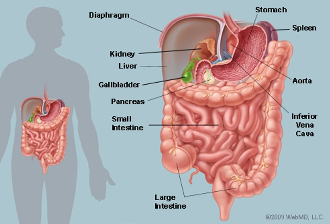
The Abdomen Human Anatomy Picture Function Parts Definition And More from img.webmd.com Abdominal wall anatomy that is clinically pertinent to the surgeon, focusing primarily on the structures of the anterior abdominal wall, will be reviewed. It's the preferred screening method for an abdominal aortic aneurysm, a weakened, bulging spot in the abdominal aorta — the major blood vessel that supplies blood to the body. Advertising on our site helps support our mission. Blood vessels of the abdomen and pelvis. Neurovasculature of the abdominal wall explore study unit superior epigastric artery: They work to carry blood away from the heart. Aortas or aortae 4) is the main blood vessel in the abdominal cavity that transmits oxygenated blood from the thoracic cavity to the organs within the abdomen and to the lower limbs. I hope this anatomy guide is helpful.
The superior vena cava is the large vein that brings blood from the head and arms to the heart, and the inferior vena cava brings blood from the abdomen and legs into the heart.
Label the abdominal blood vessels using the hints provided. These vessels are branches of the femoral artery and vein. Lateral view with the head to the right. Anatomy of shoulder 12 photos of the anatomy of shoulder anatomy of nerves in shoulder, anatomy of posterior shoulder dislocation, anatomy of right shoulder, anatomy of shoulder labrum tear, anatomy of the shoulder games, human anatomy, anatomy of nerves in shoulder, anatomy of posterior shoulder dislocation, anatomy of right. Label the biliary passages and associated structures using the hints provided. That being said, all arterial blood delivered to this region comes via branches of the abdominal aorta, and all venous blood eventually finds its way back to. The venous drainage of the abdomen is carried out by the portal venous system and the systemic venous system. Common incisions and closure techniques, and prevention and management of wound complications, are discussed elsewhere. Blood vessels of the abdomen and pelvis. Label the intestinal structures using the hints provided. The videos are done by dr. .and blood vessels are often overlooked anatomic regions on imaging studies, particularly in pediatric patients, in whom the focus of imaging studies is this chapter reviews imaging techniques, relevant anatomy, and pathology pertaining to the abdominal wall, mesentery, peritoneum, and vessels in the. Related posts of abdominal vessels anatomy anatomy of shoulder.
Posterior cardinal sinus (blue) visible after lifting up the gi tract & gonads. Nodes drain to preaortic lymph nodes in root of primary arteries of gut (celiac nodes, superior and iferior mesenteric nodes) They work to carry blood away from the heart. These vessels are branches of the femoral artery and vein. Related posts of abdominal vessels anatomy anatomy of shoulder.
Chapter 19 Blood Vessels from image.slidesharecdn.com An abdominal ultrasound is done to view structures inside the abdomen. Before lab, read through the procedure and draw diagrams of the blood vessels you will be finding and label these. Label the biliary passages and associated structures using the hints provided. Neurovasculature of the abdominal wall explore study unit superior epigastric artery: The aorta is the large artery leaving the heart. The presence of vascular loops allows surgeons to ligate individual vessels with the expectation that blood will find its way to a particular region by alternate branches. Blood, the heart and the vessels that carry blood around the body together make up the cardiovascular system. The venous drainage of the abdomen is carried out by the portal venous system and the systemic venous system.
Label the biliary passages and associated structures using the hints provided.
If you continue browsing the site, you agree to the use of cookies on this website. Introductory anatomy lab #8 slideshare uses cookies to improve functionality and performance, and to provide you with relevant advertising. This video series covers the blood vessels for anatomy and physiology ii students. Of course, recognition of the normal vascular anatomy is essential for the investigation of any abdominal vascular problem. Efferent branchial arteries injected with red latex. It is an artery, meaning that it carries blood away from the heart. (superficial epigastric vesseles labeled at center top.) details source femoral artery vein superficial epigastric vein identifiers latin arteria epigastrica superficialis mesh d019074 ta98 a12.2.16.011 ta2 4675 fma 20734 anatomical terminology [edit on. Superficial epigastric artery scheme of the femoral artery. They also take waste and carbon dioxide away from the tissues. Nodes drain to preaortic lymph nodes in root of primary arteries of gut (celiac nodes, superior and iferior mesenteric nodes) .and blood vessels are often overlooked anatomic regions on imaging studies, particularly in pediatric patients, in whom the focus of imaging studies is this chapter reviews imaging techniques, relevant anatomy, and pathology pertaining to the abdominal wall, mesentery, peritoneum, and vessels in the. Label the biliary passages and associated structures using the hints provided. Blood vessels the major vessels in the anterior abdominal wall can be divided into deep and superficial vessels (fig.
Aortas or aortae 4) is the main blood vessel in the abdominal cavity that transmits oxygenated blood from the thoracic cavity to the organs within the abdomen and to the lower limbs blood vessels labeled. Label the abdominal blood vessels using the hints provided.